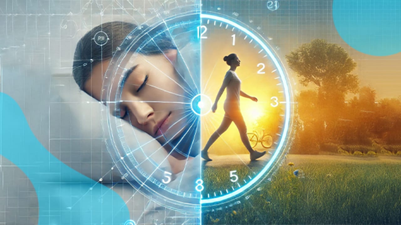


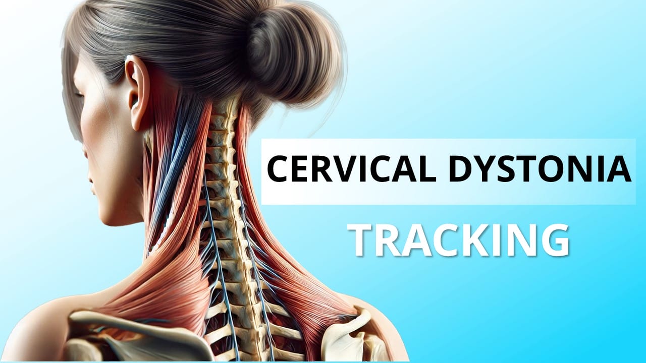
1. Introduction: The Need for Objective Cervical Dystonia Tracking
Cervical dystonia, also known as spasmodic torticollis, is a neurological disorder that causes involuntary muscle contractions in the neck, leading to abnormal head postures and movement patterns. Patients may experience twisting, tilting, or jerking of the head, often accompanied by pain and muscle stiffness. Traditionally, clinicians rely on subjective assessments, such as the Toronto Western Spasmodic Torticollis Rating Scale (TWSTRS), to evaluate symptom severity. However, these methods have limitations in tracking subtle disease progression over time.
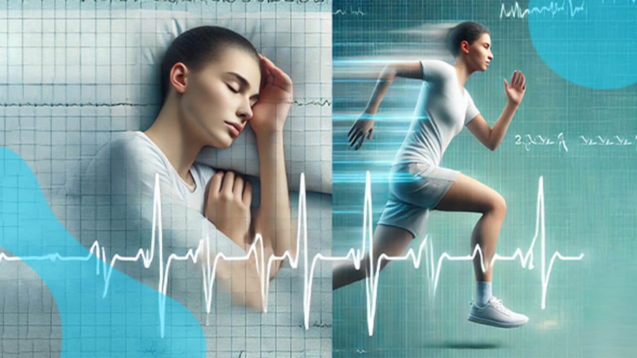
Advancements in wearable motion sensor technology now provide a way to measure cervical dystonia objectively and continuously. Accelerometers, gyroscopes, and magnetometers can track head movement, posture deviations, and tremor severity with high precision. By integrating these sensors into research and clinical practice, healthcare professionals can gain quantifiable insights into dystonia progression, ensuring better diagnosis and treatment monitoring.
2. How Wearable Motion Sensors Help in Measuring Cervical Dystonia
Wearable motion sensors have become an essential tool in movement disorder research and clinical neurology. These devices provide real-time tracking of head and neck movement, allowing for more accurate and repeatable dystonia assessments compared to traditional observational methods. Unlike clinical assessments, which can vary depending on the examiner’s expertise, motion sensors provide consistent, objective data that can be used to track changes over time.
The most commonly used motion sensors for cervical dystonia monitoring include:
- Accelerometers – Measure linear acceleration and detect head tilt and tremor.
- Gyroscopes – Measure angular velocity to capture rotational movements of the head.
- Magnetometers – Measure orientation relative to Earth’s magnetic field, ensuring long-term drift correction.
These sensors can be used independently or in combination within 9-axis motion sensors (Inertial Measurement Units – IMUs). The choice of sensor depends on the desired measurement outcome, whether it is head posture deviation, tremor characteristics, or movement smoothness.
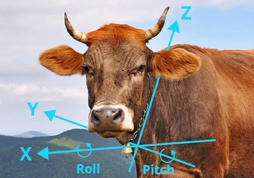
3. Using a 3-Axis Accelerometer for Cervical Dystonia Monitoring
A 3-axis accelerometer is one of the simplest and most commonly used motion sensors in wearable technology. It measures acceleration along three perpendicular axes (X, Y, and Z), allowing researchers and clinicians to analyze tilt, movement initiation, and tremor frequency. This makes it a useful tool for tracking static head posture and movement irregularities in cervical dystonia patients.
3.1 What is a 3-axis accelerometer?
A 3-axis accelerometer records how quickly an object moves in different directions. In the context of cervical dystonia, it captures head movement dynamics and detects abnormal postural changes. Since acceleration data also includes the effect of gravity, it is possible to estimate head tilt angles by analyzing the direction of gravitational force relative to the sensor.
3.2 Key Variables Measured with a 3-Axis Accelerometer
By processing raw accelerometer data, several important variables can be extracted to assess cervical dystonia severity and progression. These metrics provide insights into postural abnormalities, movement smoothness, and tremor frequency.
- Head tilt angles – Measures the degree of deviation from a neutral head position, identifying postural abnormalities such as laterocollis, anterocollis, and retrocollis.
- Movement onset and acceleration – Detects when and how fast the head moves, helping differentiate voluntary vs. involuntary movements.
- Peak acceleration – Identifies sudden, jerky movements characteristic of dystonia spasms.
- Log Dimensionless Jerk (LDJ) – A measure of movement smoothness; higher values indicate more erratic and less controlled motion.
- Power Spectral Density (PSD) for tremor analysis – Identifies dominant tremor frequency (typically in the 4–6 Hz range for dystonic tremor).
3.3 Limitations of Using Only an Accelerometer
While accelerometers provide valuable insights into movement and posture, they have limitations when used alone. A major drawback is their inability to distinguish between rotational and translational movements. This means that an accelerometer cannot differentiate whether a head tilt is caused by an actual neck movement or a shift in body posture.
Another limitation is drift and orientation tracking. Since accelerometers measure acceleration rather than position, long-term posture tracking can be inaccurate without additional data from gyroscopes or magnetometers. Furthermore, accelerometers struggle to measure angular velocity and rotational tremors, making them less effective for tracking certain dystonic symptoms compared to more advanced motion sensors.
4. Using a 9-Axis Motion Sensor for Advanced Dystonia Measurement

A 9-axis motion sensor, also known as an Inertial Measurement Unit (IMU), combines a 3-axis accelerometer, a 3-axis gyroscope, and a 3-axis magnetometer. This combination provides a complete 3D tracking system that can measure head orientation, rotational velocity, tremor frequency, and smoothness of movement. Compared to a standalone accelerometer, 9-axis sensors offer superior accuracy, better tremor detection, and the ability to track long-term posture changes in cervical dystonia patients.
4.1 What is a 9-axis motion sensor?
A 9-axis motion sensor integrates three different sensors to provide precise motion tracking:
- Accelerometer – Measures linear acceleration to detect movement onset and head tilt.
- Gyroscope – Measures angular velocity to capture head rotation, speed, and movement irregularities.
- Magnetometer – Measures orientation relative to Earth’s magnetic field, correcting drift and ensuring accurate long-term tracking.
By combining these three sensor types, a 9-axis IMU can continuously track head motion across multiple planes, making it one of the most reliable tools for monitoring dystonia severity and progression.
4.2 Key Variables Measured with a 9-Axis Motion Sensor

By fusing accelerometer, gyroscope, and magnetometer data, a 9-axis sensor can provide high-precision motion tracking for cervical dystonia assessment. The key measurable variables include:
- Absolute head orientation (yaw, pitch, roll angles) – Tracks head deviations in all three planes, allowing precise measurement of dystonic posture.
- Range of motion (ROM) – Measures how far the patient can voluntarily rotate, tilt, or flex their head.
- Peak angular velocity (°/s) – Identifies how quickly the head moves, which can differentiate normal movement from dystonic spasms.
- Angular jerk (°/s³) – Measures movement smoothness by detecting sudden changes in acceleration.
- Tremor frequency and amplitude – Captures rhythmic head oscillations and analyzes their intensity, useful for differentiating dystonic vs. essential tremors.
- Head stability index – Measures how much the head drifts when attempting to hold a steady position, indicating motor control deficits.
4.3 Advantages of 9-Axis Over 3-Axis Sensors
While a 3-axis accelerometer can provide basic movement and posture data, a 9-axis motion sensor offers superior tracking capabilities for cervical dystonia patients. The main advantages include:
- More precise head tracking – Provides full 3D motion analysis, improving accuracy in detecting dystonic movements.
- Better tremor characterization – Can distinguish between voluntary and involuntary movements using gyroscope and accelerometer fusion.
- Orientation correction – Magnetometer prevents sensor drift, ensuring reliable long-term monitoring.
- More comprehensive movement analysis – Enables tracking of posture, velocity, acceleration, and jerk in a single device.
These advantages make 9-axis motion sensors the preferred choice for clinical studies and research on cervical dystonia progression.
5. Best Practices for Sensor Placement in Cervical Dystonia
The accuracy and comfort of wearable motion sensors depend heavily on where and how they are attached to the body. Choosing the right sensor placement ensures optimal data collection while maintaining patient comfort.
5.1 Where to Attach the Sensor?
For cervical dystonia monitoring, different attachment points can provide different insights into head movement and stability. The most common sensor placements include:
- Back of the head (near the occipital bone) – The best location for tracking head posture and tremor while minimizing discomfort.
- Dual-sensor setup (head + chest) – Placing one sensor on the head and another on the upper torso allows for differentiation between head and whole-body movements.
- Neck-based or wearable integration (glasses, hats, or pendant sensors) – Embedding the sensor into everyday wearables makes long-term monitoring more comfortable and socially acceptable.
5.2 Comfort and Usability Considerations
For long-term monitoring, the sensor should be lightweight, discreet, and easy to wear without interfering with daily activities. Key considerations include:
- Non-invasive design – Headbands, clips, or adhesive patches ensure secure but comfortable sensor attachment.
- Social acceptability – Integrating sensors into glasses, hats, or clothing can help reduce self-consciousness in patients.
- Secure but flexible fit – Prevents sensor slippage while allowing full range of motion.
A well-placed sensor ensures reliable data collection while keeping patients engaged in long-term tracking studies.
6. Data Processing: How Motion Sensor Data Translates into Clinical Insights
Once data is collected from a 3-axis accelerometer or 9-axis motion sensor, it must be processed and analyzed to extract meaningful insights about cervical dystonia progression. This involves signal filtering, feature extraction, and statistical or AI-based analysis.
6.1 Signal Processing Techniques
Raw motion sensor data is often noisy and requires preprocessing to ensure accuracy. The most commonly used signal processing techniques include:
- Low-pass filtering – Removes unwanted noise and artifacts from acceleration and gyroscope data.
- Sensor fusion algorithms (Kalman filter, Madgwick filter) – Combine accelerometer, gyroscope, and magnetometer data to produce drift-free motion tracking.
- High-pass filtering for tremor detection – Isolates tremor signals (typically in the 4–6 Hz range) from other movement patterns.
6.2 Feature Extraction for Dystonia Progression Analysis
Once filtered, motion sensor data is analyzed to extract clinically relevant features. These features help quantify dystonia severity and track disease progression.
- Neck range of motion (ROM) – Tracks voluntary movement limits over time.
- Tremor analysis (Fourier Transform, Power Spectral Density) – Identifies tremor frequency, amplitude, and stability.
- Movement smoothness (log dimensionless jerk, entropy analysis) – Quantifies how erratic or controlled a patient’s movements are.
6.3 Using Machine Learning to Enhance Dystonia Tracking
Advanced machine learning algorithms are now being used to detect patterns in motion sensor data and predict disease severity or progression. These methods include:
- Supervised learning (SVM, Random Forests, Neural Networks) – Classifies dystonia severity based on sensor-derived features.
- Time-series modeling (LSTMs, ARIMA) – Predicts long-term symptom progression using past movement data.
- Automated tremor classification – Differentiates between dystonic vs. essential tremor using frequency and variability metrics.
By combining motion sensor tracking with AI-based analysis, researchers and clinicians can automate dystonia assessment and improve long-term monitoring strategies.
7. Real-World Applications and Research on Wearable Motion Sensors for Cervical Dystonia
Wearable motion sensors have become an essential tool in neurology research and clinical practice, providing an objective way to assess movement disorders like cervical dystonia. Several studies have explored the use of accelerometers, gyroscopes, and 9-axis motion sensors to track dystonic movements, tremor severity, and disease progression. These technologies not only improve clinical assessments but also open doors for at-home monitoring and remote patient tracking.
7.1 Recent Studies Using Motion Sensors for Cervical Dystonia
Researchers have tested wearable motion tracking systems in various clinical and experimental settings. Several key studies demonstrate the effectiveness of accelerometers and IMUs in dystonia assessment:
- Study on head posture tracking: Researchers attached IMUs to the back of the head to quantify postural deviations in cervical dystonia patients. The system successfully detected abnormal head tilts and provided reliable posture correction feedback.
- Study on tremor quantification: A 9-axis sensor placed on the head and chest differentiated dystonic tremor from essential tremor by analyzing tremor amplitude, frequency, and cycle variability.
- Study on movement smoothness: A machine-learning model trained on IMU-derived features accurately classified dystonic vs. non-dystonic movement based on log dimensionless jerk and tremor instability.
These studies show that wearable motion sensors are not just research tools—they are paving the way for better clinical assessments and treatment optimization.
7.2 Clinical Validation and Use Cases in Neurology Research
The application of wearable motion sensors in clinical settings has already demonstrated significant advantages:
- Objective diagnosis: Sensor-based assessments help clinicians quantify dystonic postures and tremors more accurately than visual observation alone.
- Treatment monitoring: Motion data can track the effects of botulinum toxin injections or physical therapy interventions over time.
- Home-based tracking: Wearable devices allow remote patient monitoring, reducing the need for frequent hospital visits.
Clinicians and researchers are increasingly integrating motion sensors into standard neurology workflows to provide more personalized, data-driven care for cervical dystonia patients.
7.3 Future Trends in AI-Driven Motion Analysis for Dystonia
As machine learning and AI continue to evolve, automated dystonia monitoring is becoming a reality. Emerging trends include:
- AI-powered tremor classification: Algorithms can automatically differentiate dystonic tremor from Parkinsonian tremor using sensor-derived features.
- Predictive modeling for disease progression: Time-series analysis of motion sensor data helps identify early signs of worsening symptoms, enabling proactive treatment adjustments.
- Wearable-integrated therapy guidance: Future IMU-based systems may provide real-time feedback to patients, helping them practice corrective exercises and improve movement control.
By integrating motion sensors with AI, cervical dystonia tracking will become more accessible, scalable, and precise, revolutionizing patient care.
8. Conclusion: The Future of Wearable Sensors in Dystonia Management
The use of wearable motion sensors in cervical dystonia research and clinical practice is transforming how neurologists diagnose, track, and manage the condition. Traditional clinical assessments rely on subjective ratings, but accelerometers, gyroscopes, and 9-axis motion sensors provide real-time, quantifiable movement data that enhances diagnostic precision.
A 3-axis accelerometer is useful for detecting posture deviations, movement onset, and tremor frequency, while a 9-axis motion sensor offers advanced tracking of head orientation, rotation speed, and movement smoothness. By strategically placing sensors on the head, neck, or chest, clinicians can obtain high-quality movement data without disrupting patient comfort.
With advances in machine learning and AI-powered motion analysis, wearable sensors are becoming a cornerstone of dystonia research and treatment optimization. As more clinical studies validate these methods, we can expect motion-tracking technology to become a standard tool for dystonia management in both hospitals and home settings.
Key Takeaways:
- AI-powered motion analysis will further enhance dystonia tracking, enabling predictive models for disease progression.
- Wearable motion sensors provide an objective way to track cervical dystonia symptoms and progression.
- A 3-axis accelerometer measures basic movement patterns, while a 9-axis sensor provides advanced tracking of tremor, posture, and movement smoothness.
- Proper sensor placement ensures reliable data collection, improving both clinical assessments and at-home monitoring.
Call to Action
For more guidance on selecting the best device for your study, explore You may book a video call with our expert or ask for a quote.

Frequently Asked Questions
What is cervical dystonia, and how does it affect movement?
Cervical dystonia is a neurological disorder that causes involuntary muscle contractions in the neck, leading to abnormal head postures, tremors, and movement difficulties. It can result in pain and stiffness, impacting daily activities.
How can wearable motion sensors help track cervical dystonia?
Wearable motion sensors, such as accelerometers, gyroscopes, and magnetometers, provide real-time tracking of head movement, posture, and tremor severity. They offer objective, consistent data that help monitor disease progression and treatment effectiveness.
What are the benefits of using a 9-axis motion sensor over a 3-axis accelerometer?
A 9-axis motion sensor combines accelerometers, gyroscopes, and magnetometers, providing precise 3D tracking of head orientation, movement smoothness, and tremor patterns. It offers superior accuracy compared to a 3-axis accelerometer, which primarily measures linear acceleration and tilt.
Where should wearable sensors be placed for cervical dystonia monitoring?
The optimal placement for motion sensors depends on the measurement goal. The back of the head (occipital region) is ideal for tracking posture and tremor. A dual-sensor setup (head + chest) helps differentiate head movements from body shifts. Wearable integrations like glasses or hats improve long-term comfort.
Can AI and machine learning improve cervical dystonia assessment?
Yes, AI algorithms can analyze motion sensor data to classify dystonia severity, predict symptom progression, and differentiate between different movement disorders. Machine learning models enhance tracking accuracy and enable automated, data-driven assessments.